Akoya
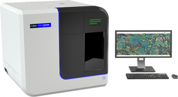
PhenoImager HT
Log in for pricing
As part of our PhenoImager™ translational solution, multispectral imaging on the PhenoImager HT can be applied across a whole slide using the 6-plex, 7 Color Opal Polaris reagent kit. This enables the biology to be explored at multiple scales, from cell-to-cell interactions to the macroscopic tissue architecture. The whole slide multispectral imaging capability creates a simpler and more robust workflow as fields of view do not need to be selected eliminating selection bias. You also retain a whole slide record, no re-scans required, so that you can easily re-analyze imagery as new understanding emerges. Tissue sections or TMAs can be labeled with immunofluorescent (IF) or immunohistochemical (IHC) stains such as Opal™, or with conventional stains such as H&E and trichrome. When using IF or IHC stains, multiple proteins can be measured on a per tissue, per cell, or per cell compartment (e.g. nuclear, cytoplasmic) basis – even when signals are spectrally similar, are located in the same cellular compartment or are obscured by autofluorescence.
As part of our PhenoImager™ translational solution, multispectral imaging on the PhenoImager HT can be applied across a whole slide using the 6-plex, 7 Color Opal Polaris reagent kit. This enables the biology to be explored at multiple scales, from cell-to-cell interactions to the macroscopic tissue architecture. The whole slide multispectral imaging capability creates a simpler and more robust workflow as fields of view do not need to be selected eliminating selection bias. You also retain a whole slide record, no re-scans required, so that you can easily re-analyze imagery as new understanding emerges. Tissue sections or TMAs can be labeled with immunofluorescent (IF) or immunohistochemical (IHC) stains such as Opal™, or with conventional stains such as H&E and trichrome. When using IF or IHC stains, multiple proteins can be measured on a per tissue, per cell, or per cell compartment (e.g. nuclear, cytoplasmic) basis – even when signals are spectrally similar, are located in the same cellular compartment or are obscured by autofluorescence.


