Akoya
Opal 6-Plex Detection Kit (Whole Slide Imaging) - For Manual Workflow
Log in for pricing
Formerly Known as: Opal Polaris 7 Color Manual IHC Detection Kit
The Opal 6-Plex Manual Detection Kit - for Whole Slide Imaging is part of the PhenoImager whole slide workflow and contains:
- 6 reactive fluorophores (Opal 480, Opal 520, Opal 570, Opal 620, Opal 690, and Opal 780)
- 10X Spectral DAPI
- DMSO
- 1X Plus Manual Amplification Diluent
- 1X Antibody Diluent/Block
- Opal Anti-Ms + Rb HRP
- AR6 Buffer
Users should source primary antibodies and other workflow reagents separately. The Opal 6-Plex Manual Detection Kit - for Whole Slide Imaging was optimized for use on the Vectra® Polaris™ quantitative pathology imaging system. Kits contain enough reagents to stain up to 50-slides at the recommended 1:100 dilution ratio.
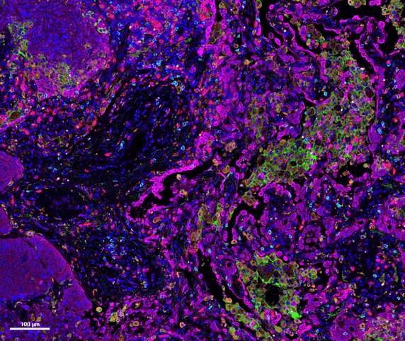
What is Opal? Opal Multiplex IHC Kits make multiplex methods accessible to researchers who works with standard immunohistochemistry (IHC). The Opal method allows use of any number of unlabeled primary antibodies from the same species in multiplexed tissue assays, with no fear of cross-reactivity. Antibodies for simultaneous IHC may now be selected based on performance, rather than species. The method works with formalin fixed paraffin-embedded (FFPE) tissue and is compatible with the standard research IHC workflow in your lab.
Opal provides researchers with the tools to interrogate multiple pathways while retaining context provided by tissue images. This approach provides information not available from alternative techniques like flow cytometry or analysis of single markers in serial sections.
Opal will enable you to:
Measure up to six tissue biomarkers at once
Use the best primary antibodies together in multiplex panel, with no species-based crosstalk
Retain spatial cellular context that is lost in flow cytometry
Confirm single cell co-expression for many biomarkers in one tissue section
Get more information while conserving precious tissue.


